The Causes | MR-Sim
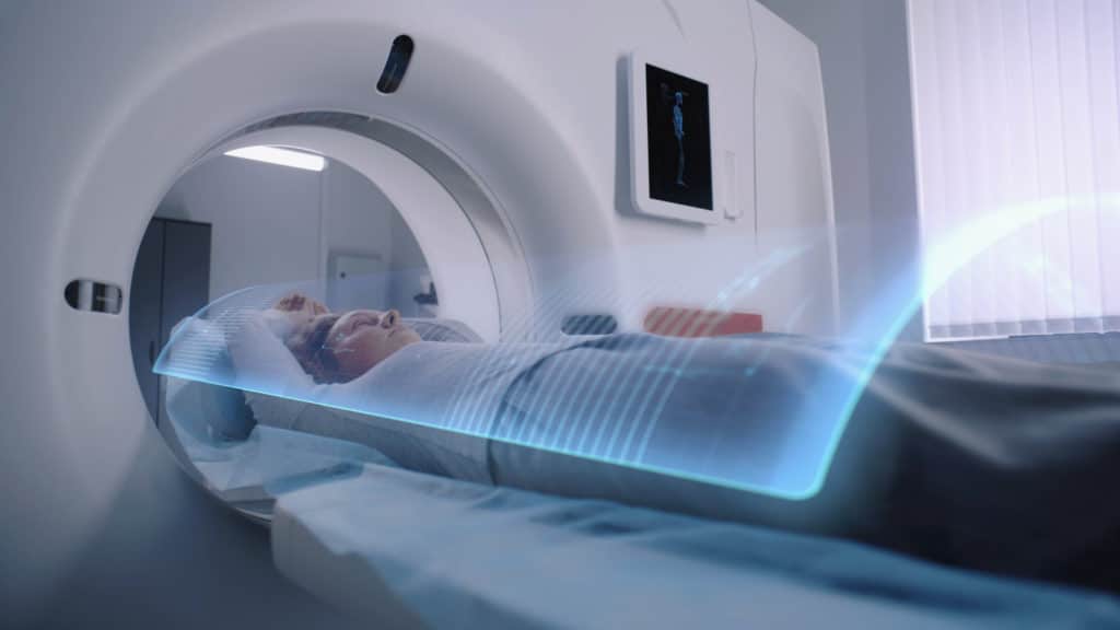
What is an MR-Sim?
Medical professionals have long used Computed Tomography (CT) technology to see inside the human body to find and target cancer tumours with radiation. CT technology takes X-ray images of the subject from different angles and uses computer processing to create images of soft tissues in a patient’s body.
Magnetic resonance imaging (MRI) uses a magnetic field and radio pulses to create pictures of organs and soft tissues. A magnetic resonance simulator (MR-Sim) is similar to an MRI, but unlike a standard MRI, the MR-Sim is specifically designed for the planning and preparation stages of radiation treatment, enabling more accurate targeting of tumours.
An MRI Scan can be seen on the left, and a CT scan on the right. The resolution of the MR scan is much higher, making it way easier to see the cancerous area.

Enhanced Imaging for Better Diagnosis
The MR-Sim provides high-resolution images of cancerous areas, allowing doctors to see tumours and their edges more clearly and precisely than with a CT scan.
Improved Precision in Treatment Planning
These clear, detailed images enable more precise planning for radiation therapy, ensuring that treatment is accurately targeted at cancerous areas while avoiding healthy tissue.
More Effective Radiation Delivery
With improved imaging and planning, higher doses of radiation can be directed specifically at tumours, significantly reducing the risk of damaging surrounding healthy tissues and organs.
Less chance of Recurrence
With the ability to see considerably smaller cancer cells on the edges of tumours and target them as well, there is a reduction in the chance of the cancer returning.
Shorter Treatment and Recovery Times
By using higher doses of radiation and minimizing harm to healthy cells, the MR-Sim reduces the overall treatment window, leading to faster recovery times.
Increased Capacity for Patient Care
The efficiency of the MR-Sim allows medical professionals to treat more patients, enabling quicker access to care and reducing waiting times for those in need of treatment.
What Types of Cancer will an MR-Sim Help to Treat?
- Breast Cancer
- Prostate Cancer
- Lung Cancer
- Liver Cancer
- Head and Neck Tumours
What is a Linac MR?
The Linac MR is a groundbreaking invention conceived here in Alberta with Cure Cancer Foundation funding. Dr. Gino Fallone had the idea for the device, which was initially thought impossible. The Linac MR incorporates a linear Accelerator (linac) and a Magnetic Resonance Imaging (MRI or MR), which were believed to be incompatible until Dr. Fallone had a breakthrough idea on how to combine them.
Previously, patients were scanned with an MRI to find tumours, then moved to a Linac for radiation therapy. By combining both machines, doctors can do both at the same time, ensuring the precise delivery of radiation. This new cancer treatment method significantly improves the accuracy of radiation therapies. The Linac MR has the potential to revolutionize how cancer is treated.
An MR-Sim will allow for better planning, making the Linac MR a more effective cancer treatment tool.
How Can an MR-Sim Help to Treat Cancer?
Higher-Resolution Imaging
Sadly, you can’t treat what you can’t see. The MR-Sim provides much clearer images of tumours, allowing doctors to define the edges of cancers more precisely.
Tumours located between the neck and pelvis, such as breast and bowel cancers, are very difficult to treat because the cancer cells can shift positions. For example, when a cancer patient breathes, or their bladder or bowels fill up, the cancerous tissue moves as well. This movement makes it difficult to precisely target the cancer cells with radiation without also affecting vital organs, such as the heart in breast cancer patients or the bowel and other nearby areas in prostate cancer patients. The MR-Sim offers very high-resolution images, which will allow doctors to plan radiation treatments with the Linac MR better. Thus making it much easier to deliver radiation doses to tumours while avoiding healthy cells.
Better Planning
Radiation therapy, like treating an infection with penicillin, requires careful dosing to avoid harmful effects. Just as penicillin is administered in smaller, manageable doses, radiation treatments must be spread out to minimize damage to surrounding healthy tissue.
The MR-Sim will make planning treatments easier by offering much higher resolution images of a cancerous area and, therefore, much better looks at the tumours. This will allow doctors to hit cancer with higher doses of radiation precisely.
This planning will reduce the number of treatments needed, potentially lowering the number of radiation therapy sessions from around thirty to just a few, thereby significantly shortening recovery times for patients.
Reducing Damage to Healthy Cells
The MR-Sim will significantly reduce damage to healthy cells by providing highly detailed imaging that doctors can use to distinguish cancerous cells from healthy tissue. This will help ensure that radiation is focused solely on cancer cells while minimizing exposure and reducing collateral damage to surrounding tissue. The MR-Sim will dramatically reduce the recovery time and long-term effects of treatment for cancer patients.
More Patients Treated
At the Cross Cancer Institute, machine time is a valuable resource. The MR-Sim will help reduce the treatment time for each patient, allowing more patients to receive care faster. This is particularly beneficial for individuals from rural Alberta or the Northwest Territories, as it reduces the time they need to spend away from their homes for treatment.